
ONGOING PROJECTS
- Development of Remote Radiation Detection Imaging System Mounted on a Drone
- High Resolution SPECT using Variable Pinhole Collimator
- Positioning Algorithm for CZT Virtual Frisch-grid Detector
- Multi-purpose Super-resolution Gamma Detector
- Multi-pinhole SPECT
- Low Profile Light Guide using Diffusion Film
- Advanced Reconstruction for Radiation imaging (ARRA)
COMPLETED PROJECTS
- Reconstruction of Dose Distribution in In-beam PET for Carbon Theraphy
- Image Registration for Breast Cancer Study
- High Energy Collimator Design for I-131
- Plasma-Display-Panel based X-ray Detector (PXD)
- CCD based Gamma Camera
- Abdomen Registration for PET/CT and MR images
- Performance Optimization by Patient Dose Analysis and Image Quality Assessment in CT Fluoroscopy
- Cone-beam based system matrix for a pixelated SPECT detector
- Development of Time-of-Flight Method for Improvement of Signal-to-Noise Ratio
High Resolution SPECT using Variable Pinhole Collimator
Introduction
Single-photon emission computed tomography (SPECT) is one of the most widely used medical imaging techniques in the field of nuclear medicine. The two major components that determine the quality of a SPECT image are the collimator and the scintillation detector. The collimator confines the direction of the incident gamma rays that can arrive at the detector, so that the direction of the emitted photon can be estimated. Different types of collimators are used in SPECT based on the application at hand. In preclinical imaging, pinhole collimators, which have a higher spatial resolution than other collimators, are used frequently. However, one of the drawbacks of the pinhole collimator is its low sensitivity, which occurs by virtue of its intrinsic design. In an ideal gamma imaging system, the sensitivity and spatial resolution of the knife-edge pinhole collimator is determined by the acceptance angle(θ), diameter(r), and distance(d) between the pinhole and the object, as illustrated in Fig. 1.
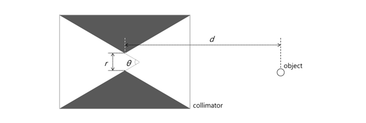
Fig. 1. Pinhole collimator and its design parameters.
In general, the design parameters of a pinhole collimator are predetermined to acquire the gamma image of the whole object. The center of the pinhole aperture in SPECT is aligned to the center of the gantry rotation as well. As a final step, the predesigned pinhole collimator, which is drilled in a single tungsten block, is placed in the gamma imaging system. Once the pinhole is manufactured, the acceptance angle, diameter, and distance from the object cannot be changed. Therefore, the conventional pinhole collimator based SPECT system can reconstruct the whole body gamma image only for an object that has the same acceptance angle, diameter and distance from the pinhole, for every rotation angle. Even though the region-of-interest(ROI) is specified as a target organ, because the field-of-view(FOV) of conventional pinhole SPECT is set to the whole object, the resulting image of the target organ can have poor sensitivity and spatial resolution. Here, we propose a novel variable pinhole collimator whose parameters are flexible. In this collimator, the shape of the pinhole can be optimized to the ROI for each rotation angle, resulting in an improvement in the sensitivity and spatial resolution of the target oriented SPECT. In this study, the concept of a variable pinhole is introduced and the performances of the proposed SPECT are also presented.
Materials and Methods

Fig. 2. (a) Cross-sectional diagram of the variable pinhole collimator, which has been designed by piling tungsten layers having different apertures. (b) Different apertures which are drilled into a tungsten sheet. (c) The movements of stacked tungsten sheets are guided by motor controlled sliding units.
The proposed variable pinhole collimator, modeled on the conventional pinhole by piling several tungsten layers of different apertures, is shown in Fig. 2 (a). These apertures are drilled into thin tungsten sheets, as displayed in Fig. 2 (b) and the movement of each tungsten sheet is controlled by its designated motor driving unit, to align the optimal aperture of each layer with the pinhole center, as shown in Fig. 2 (c). If each tungsten sheet is thin enough and the number of layers large enough, the diameter and acceptance angle of the variable pinhole can imitate that of the conventional pinhole. Since the FOV is predetermined in conventional SPECT, the size of the reflection on the scintillation detector, which has been formed by the pinhole, is predetermined as well, to fully cover the area of the scintillation detector. In contrast to this, in the proposed target oriented SPECT, the size of the ROI(x) and the distance of the collimator from the object(d1) can be varied for each target organ and rotation angle. Because the acceptance angle can be modified in the variable pinhole collimator, the size of the reflection image(y) for every rotation angle can be ascertained to cover the entire area of the scintillation detector, by varying the distance of the pinhole from the scintillation detector(d2), for each rotation.

Fig. 3. Schematic diagram depicting the movements of the conventional SPECT system (a) and the target oriented SPECT system (b), through a complete rotation.
Fig. 3 shows conventional SPECT system (Fig. 3 (a)) and the proposed target oriented system (Fig. 3 (b)). The proposed target oriented SPECT system consists of the variable pinhole collimator and several actuator units to translocate the collimator and scintillation detector for each rotation angle, to maximize the sensitivity and spatial resolution of the resulting image.
Results
To compare the performance of conventional pinhole collimator based SPECT and the proposed variable pinhole collimator based target oriented SPECT, a simulation study was conducted using GATE V6.2 on the cluster. First, we optimized the thickness of each tungsten sheet. In this simulation, a collimator made of 4-mm-thick tungsten, with an acceptance angle of 60˚ and a diameter of 0.4 mm was employed and the scintillation detector consisted of a 50 ×50 × 5 mm GAGG crystal. To measure the spatial resolution of the entire area, three ideal point sources (Tc-99m) were placed at intervals of 2 mm, from the center to the edge of the FOV.
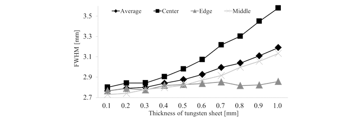
Fig. 4. FWHM of the variable pinhole collimator, for each thickness of the tungsten sheet.
Fig. 4 shows the results of the full-width at half maximum (FWHM) for each thickness of the tungsten sheet. As the thickness of the tungsten sheet is increased, the spatial resolution is reduced. From this study, the thickness of the tungsten sheet, that yields an FWHM similar to that of a conventional knife-edge pinhole (2.8 mm), is determined to be 0.4 mm. For a quantitative measure of the performance, we compared the sensitivity and spatial resolution of the reconstructed images from both conventional SPECT and the proposed target oriented SPECT. In order to do this, a 10 mm diameter of a micro-Derenzo phantom was simulated. This digital phantom was misaligned by 8 mm along the x-axis, from the center of a 30 mm diameter water phantom.

Fig. 5. Reconstructed image from (a) conventional pinhole SPECT, and (b) variable pinhole SPECT.
Fig. 5 shows the reconstructed image of the phantom, which has been obtained using the 3D maximum-likelihood expectation maximization (MLEM) algorithm. While hot rods of diameter 0.5 mm can be distinguished in the image from the proposed system, nothing is discernible in the conventional SPECT image. The sensitivity of the proposed system is higher by about 209% than the conventional pinhole system.
Conclusion
In this study, we designed a novel pinhole collimator for SPECT and showed preliminary results of target oriented imaging through a simulation study. Currently, we are pursuing strategies to realize the proposed system, in order to develop high sensitivity and high resolution preclinical SPECT.
Participating Researchers