
ONGOING PROJECTS
- Development of Remote Radiation Detection Imaging System Mounted on a Drone
- High Resolution SPECT using Variable Pinhole Collimator
- Positioning Algorithm for CZT Virtual Frisch-grid Detector
- Multi-purpose Super-resolution Gamma Detector
- Multi-pinhole SPECT
- Low Profile Light Guide using Diffusion Film
- Advanced Reconstruction for Radiation imaging (ARRA)
COMPLETED PROJECTS
- Reconstruction of Dose Distribution in In-beam PET for Carbon Theraphy
- Image Registration for Breast Cancer Study
- High Energy Collimator Design for I-131
- Plasma-Display-Panel based X-ray Detector (PXD)
- CCD based Gamma Camera
- Abdomen Registration for PET/CT and MR images
- Performance Optimization by Patient Dose Analysis and Image Quality Assessment in CT Fluoroscopy
- Cone-beam based system matrix for a pixelated SPECT detector
- Development of Time-of-Flight Method for Improvement of Signal-to-Noise Ratio
Feasibility Study of a Lens-coupled Charge-Coupled Device (CCD) Gamma Camera
Introduction
Preclinical gamma imaging is one of the most valuable methods in medical imaging. A sub-millimeter resolution detector is essential for improving the performance of gamma cameras for small animals. Conventional detectors usually consist of photomultiplier tubes (PMTs) coupled to scintillators. While the PMT produces good energy resolution, the spatial resolution is poor compared to those of other sensors. Charge-coupled devices (CCDs) are widely used as detectors in various digital imaging systems. In medical imaging systems, the CCDs have been applied to X-ray fluoroscopy and dental radiographic systems due to their compactness, light weight, high spatial resolution, and low cost. In gamma imaging, the intrinsic light signal has a very low intensity; thus, gamma cameras require high sensitivity, and low noise. The major drawback of CCDs for gamma-ray detection is their intrinsic noise, which is mainly affected by the dark current noise. A Peltier cooler directly coupled to the CCD surface reduces the temperature of the CCD to adequately suppress dark current noise. These enhanced noise-suppressing techniques increase the feasibility of a CCD-based gamma camera. In this study, we built a cost-effective CCD based gamma camera. We designed a prototype detector with a pinhole collimator, a CsI(Tl) columnar scintillator, two optically-coupled lenses (one micro-lens for short object distances, and one wide angle lens to enlarge the field of view (FOV)), and a conventional Peltier-cooled CCD, then, we measured the performance of the proposed detector. We investigated the thickness of the scintillator using Monte Carlo simulation and experimental results. We also measured the linearity of the sensitivity and the spatial resolution with a full width at half-maximum of the gamma-ray line source image. The MTF was also explored. The properties investigated through this prototype system demonstrated its feasibility for small FOV gamma imaging at low cost.
Materials and Methods
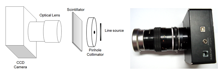
Fig. 1. Geometry of the prototype system (left) and lens coupled CCD camera module (right).
We designed and implemented a prototype CCD gamma camera system. An overview of the system is illustrated in Fig. 1 (left). A pinhole collimator was employed to form an image of a radioactive object on the columnar scintillator. Converted visible light from the scintillator was focused on the Peltier-cooled CCD surface through two optically-coupled lenses. The external light from outside the system was cut off by using a developed dark box housing covered with black PVC foam sheets. A CCD is a photoelectric semiconductor with compact size, light weight, and high spatial resolution. The size of the sensor used in this study is small (864 mm² for a full-frame CCD), and the price was reasonable for use as a general mobile cameras. Even though CCDs have various advantages compared to other light detectors, they were not recognized as appropriate detectors for gamma imaging because of their poor SNR at a low count rates. This problem was mainly caused by dark current which is primarily affected by the CCD surface temperature. Recently, a Peltier-based cooling system was developed, thus, the temperature of the detector was lower and the dark current was significantly reduced. To maximize the light collection efficiency from the scintillator to the detector, we explored various coupling methods. The use of an optical lens is a useful coupling method for cost-effective systems. To change the FOV, we need only change the lens with a valid specification. Even though the light collection efficiency was lower than that of the taper method, the price and the performance of the optical lens are reasonable. In this study, we used a micro-lens because it has good light yield and a short focal length (Fig. 1 (right)). The FOV size of this micro-lens coupled system is about 2 cm x 1.34 cm at an object distance of 35 cm. To increase the FOV size to 4 cm x 2.68 cm, which is compatible with the scintillator size, we coupled the micro-lens to a wide-angle lens.
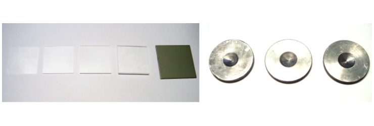
Fig. 2. Different types of scintillators (left) and pinhole collimators customized for the prototype systems (right).
Since the CCD used in this study showed a maximum quantum efficiency at around 600 nm, we chose a CsI(Tl) scintillator, which has the nearest emission wavelength. In our prototype detector, the thickness of the scintillator was one of the major parameters that affected the sensitivity of the system. To determine the optimal scintillator thickness, Monte Carlo simulation (GATE6.0) was used. The experimental sensitivity results versus the different scintillator thicknesses (0.3, 1, 2, and 4 mm) were compared to the simulation results. A 0.6 mm-thick columnar scintillator was used to obtain a high resolution image. Four types of non-columnar scintillators and a 0.6 mm-thick CsI(Tl) columnar scintillator were used in this experiment as shown in Fig. 2 (left). We designed three types of pinhole collimators dedicated to this gamma imaging system. To determine the optimal pinhole size associated with sensitivity and spatial resolution, we designed each collimator with a 0.5-, 1-, and 2-mm pinhole (Fig. 2 (right)). The collimators were made of 5-mm-thick lead, and the distance from the pinhole focal spot to the scintillator surface was set to 2 cm. The distance from the focal spot to the object was set to 1 cm to increase the spatial resolution.
The prototype CCD gamma camera was developed, and the performance experiments of the system were performed. In this prototype system, most gamma rays from the radiation sources were collected at the pinhole collimator. Only the gamma-rays within the angle range of the pinhole edge reached will reached the scintillator and be converted to visible light. This light will be collected by the CCD through two optically-coupled lenses. To decrease the dark current of the sensor, we set the temperature of the CCD to 0 celsius by using the Peltier. Two optical lenses were coupled to increase the light yield and obtain an adequate FOV. We set the aperture of the lens to F2.8 to maximize the light yield and the focal point on the surface of scintillator. We used 4 cm x 4 cm CsI(Tl) scintillators with thicknesses of 0.3, 1, 2, and 4 mm to determine system sensitivity for each scintillator's thickness. We employed a CsI(Tl) columnar scintillator (4.8 cm x 4.8 cm with 0.6 mm thickness) to obtain the MTF and line source image. The sensitivity for the each scintillator was measured with a point source (Co-57, 0.1 mCi) which was located at the center of the scintillator surface. The line source image used to obtain FWHM was taken through a 1 mm diameter the pinhole collimator with a 1.7 mCi Tc-99m radiation source inside the capillary tube (1.1 mm inner diameter). To determine the dose-to-pixel value conversion function, we measured the dose from an X-ray generator with a 500-cc ion chamber. Each CCD pixel value, from 0.2 mAs to 1 mAs of the 70-kVp X-rays was used to determine the conversion function. With these results, the MTF of the prototype system was measured using a 0.01 mm tungsten slit.
Results

Fig. 3. Simulation results of the energy deposited in the scintillator and the experimental pixel value for various scintillator thicknesses (left) and Linearity test results for the prototype system (right).
The sensitivity for various thicknesses of the scintillator was compared using the Monte Carlo simulation (GATE 6.0). Fig. 3. (left) shows the simulation and the experimental results. As the results show, we found that more gamma rays interacted inside the scintillator until its thickness was smaller than 10 mm. Although thicker scintillators showed good sensitivities, the manufacturing process used for columnar-type scintillators does not support scintillators thicker than 1 mm. Thus, we adopted a 600 micrometer-thick columnar scintillator to obtain an image with this prototype system. The difference between the simulation and the experimental results was mainly caused by the light conversion efficiency, which is dedicated by the purity of the scintillator. The linearity of the radiation sensitivity of the prototype system was tested. Fig. 3. (right) shows the linearity of the prototype system, which represents the relationship between the exposed dose and the pixel value. The Figure demonstrates excellent linearity for prototype system.

Fig. 4. Tc-99m source, 1.7 mCi, inside the 1.1-mmdiameter capillary tube.
The spatial resolution of the system was measured using a Tc-99m line source. The gamma image of a line source with a 1.1-mm inner diameter is shown in Fig. 4. The size of the collimator hole was 1 mm, the thickness of columnar scintillator was 0.6 mm, the exposure time was 20 min, and the activity of the radiation source was 1.7 mCi. With this image, we measured the FWHM of the 1.1-mm line source image, which was 2.85 mm.

Fig. 5. Line spread function (left) and MTF(right).
The MTF was measured with a 0.01 mm tungsten slit. In this experiment, we used a 50-kVp X-ray source, which is compatible to the slit size. Figure 8(left) shows the line spread function, and Fig. 8(right) shows the MTF of the prototype system. The FWHM of 0.01 mm-slit line spread function was measured to be 0.37 mm.
Conclusion and Discussion
The objective of this study was to investigate the feasibility of a CCD-based gamma camera for preclinical studies. We designed a compact and low cost prototype system with high sensitivity and spatial resolution. To achieve a high spatial resolution image, we combined a Peltier-cooled CCD with a CsI(Tl) columnar scintillator. For high light collection efficiency and adequate FOV, a micro-lens attached to a wide angle lens was used to deliver light from the scintillator to the CCD. To block light photons from outside the system, we built a dark box housing covered with black PVC foam sheets. The performance of the prototype system was measured. We obtained the sensitivity through various scintillator thicknesses, the linearity representing the relationship between the dose and the pixel value, and the spatial resolution of a line source image. The linearity of the sensitivity was outstanding, and would be very useful for applications that need to measure the radiation activity. The 1.1 mm gamma-ray line source image with the pinhole collimator and columnar scintillator showed a 2.85-mm FWHM. Moreover, the MTF of this system was measured with a 0.01mm tungsten slit. Our investigation of this prototype system demonstrated its feasibility for use as a high performance low cost small animal gamma camera.
Participating Researchers