
ONGOING PROJECTS
- Development of Remote Radiation Detection Imaging System Mounted on a Drone
- High Resolution SPECT using Variable Pinhole Collimator
- Positioning Algorithm for CZT Virtual Frisch-grid Detector
- Multi-purpose Super-resolution Gamma Detector
- Multi-pinhole SPECT
- Low Profile Light Guide using Diffusion Film
- Advanced Reconstruction for Radiation imaging (ARRA)
COMPLETED PROJECTS
- Reconstruction of Dose Distribution in In-beam PET for Carbon Theraphy
- Image Registration for Breast Cancer Study
- High Energy Collimator Design for I-131
- Plasma-Display-Panel based X-ray Detector (PXD)
- CCD based Gamma Camera
- Abdomen Registration for PET/CT and MR images
- Performance Optimization by Patient Dose Analysis and Image Quality Assessment in CT Fluoroscopy
- Cone-beam based system matrix for a pixelated SPECT detector
- Development of Time-of-Flight Method for Improvement of Signal-to-Noise Ratio
A Development of Image Reconstruction Algorithm for the Multi-Pinhole Collimator of I-131 Based Gamma Camera
Introduction
I-131 has been widely used for radiotherapy owing to its relatively long half-life of 8.02 days and its simultaneous emission of high gamma and beta rays. This isotope is used in thyroid postoperative adjuvant therapy and on metastatic diseases. In addition, it can be used to generate a post treatment image to verify the follow-up results. Despite these advantages, collimator penetration, due to the high energy peaks of I-131 (364 keV), fundamentally degrades the quality of I-131 imaging. Particularly, the problem has a more significant in preclinical research because of the small rodent size and required high resolution. Recently, there have been several new applications of preclinical single photon emission computed tomography (SPECT). The preclinical SPECTs provide energy range conditions, which were set to various radioisotopes in the preclinical field. Although the applications have been developed, high energy peak scatter, and penetration issues were still critical issues. It is highly important to minimize the penetration and scatter, and to preserving the main peak sensitivity by designing an optimized I-131 pinhole collimator for small animal SPECT. Penetration can affect the spatial resolution and non-uniformity of the sensitivity distribution. For these reasons, the new pinhole aperture design needs specific part for reducing penetration. The channel insert design for the pinhole has been investigated to reduce penetration. To achieve better collimator resolution, the pinhole size and acceptance angle need to be optimized together for high sensitivity. The aims of this study are to design a high energy pinhole collimator for the I-131 small animal SPECT and to model the structure by Monte Carlo simulation, and finally to validate the results by the sensitivity and the spatial resolution. We performed simulation by GATE V6.0 (Geant4 Application for Emission Tomography), which is an advanced open-source software which dedicated to numerical simulation in medical imaging and radiotherapy.
Materials and Methods
1. Collimator design
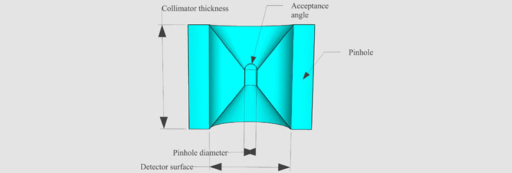
Fig. 1. Pinhole design parameters.
Fig. 1 shows several parameters used to design pinhole collimators. There are some main parameters required to design collimator: collimator thickness, pinhole diameter, acceptance angle etc. Collimator thickness influences the amount of the attenuation when the photons from radiation source move to detector through the collimator. Pinhole diameter has an effect on sensitivity and spatial resolution. The acceptance angle affects the size of FOV(Field Of View) and the sensitivity. We modeled various design of collimator with different values of each parameter using GATE v6.1(Geant4 Application for Emission Tomography).
2. Image reconstruction for SPECT data
SPECT is the system that makes tomographic image with the projection images at each angle using rotating gamma imaging device. We implemented the maximum likelihood expectation minimization(MLEM) algorithm to get reconstructed SPECT image. This algorithm iteratively computes expected back-projected image and projection data to get converged reconstruction image. Fig. 2 is a block diagram which shows the flows of the algorithm.

Fig. 2. Block diagram of MLEM algorithm.
When the algorithm computes the two kinds of image(projection, back-projection), it is needed to calculate the detecting probability between image pixels and detector bins. The results of the calculation is called system matrix. There are three main methods of calculating system matrix as following figure. We developed unmatched system matrix calculation which used ray-driven method for projection and pixel-driven method for back-projection to enlarge the efficiency of calculation accuracy per computing time.
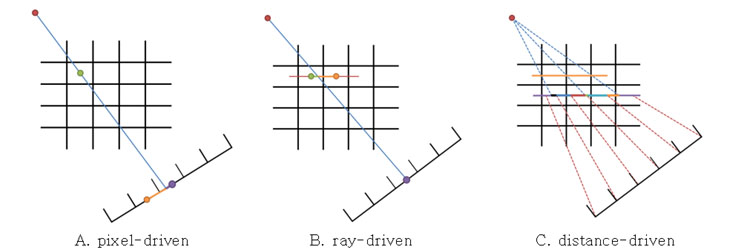
Fig. 3. System matrix calculation methods.
Pixel-driven method: Calculate the contribution of the LOR(line of response) from the center of the each image pixel to detector bin.
Ray-driven method: on the contrary to the pixel-driven method, calculate the contribution of the LOR from the center of the each detector bin to image pixel.
Distance-driven method: Calculate the contribution of the LOR on the virtual plane made with detector bins and image pixels.
3. Projection image evaluation

Fig. 4. Simulation condition for multi-pinhole collimator.
We modeled the multi-pinhole collimator for getting projection image and evaluation. Fig 4. shows the GATE simulation condition. Detector material was NaI, collimator material was tungsten with 15mm thickness to sufficient attenuation and the phantom in image shows the range of FOV. The X, Y, and Z position values in the interaction data sets contained the position of interaction on the detector. 313.2 × 193.2 (mm2) planar projection images were generated as gateSingles.dat files from GATE simulation. Gaussian blurring filter with a standard deviation of D0 for smoothing line profile described by Eq. 1, was applied to the frequency domain of the planar images.
Results
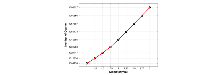
Fig. 5. Detected counts from 1.0mm to 3.0mm pinhole diameter.
Fig. 5 shows the number of the detected photon through the a pinhole collimator whose diameter is from 1.0mm to 3.0mm. As pinhole diameter increases, the count also increases but the spatial resolution decreases.

Fig. 6. Projection image with 5mm sphere source(left) and profile of the projection image(right).
Fig. 6 shows the projection image at a single angle and the line indicates where we get image profile. With 1.5 mm pinhole diameter and 5mm sphere source simulation, the FWHM for each pinhole was 24.6mm, 19.3mm, 22.1mm.
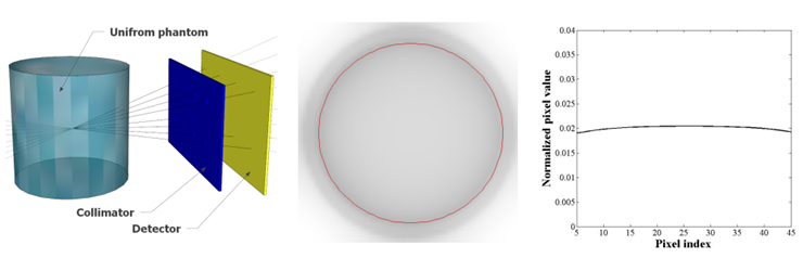
Fig. 7. Simulation condition for projection data(left), reconstructed image with uniform phantom(middle), profile of the image(right).
Fig. 7 shows the reconstruction results. The left figure is the simulation condition to getting projection data for reconstruction input, the middle figure is the reconsturction output whose FOV is inside of red circle and the right figure shows profile of the reconstruction results.
Conclusion
In this study, we designed the multi-pinhole collimator for I-131 source and physically simulated the pinhole structure. And we implemented MLEM reconstruction algorithm with unmatched system matrix calculation for efficient accuracy and computing time. Projection images were generated from gamma interactions at the scintillation crystal in the simulation. We determined the acceptance angle in order to maximize the true count rate, which was found to correspond to the crossing-point of the plots of sensitivities of the penetration and scatter photon count rates. A pinhole diameter of 1.5 mm produced a minimum FWHM value for the projection images. A collimator thickness of at least 20 mm was required to reduce the penetration photon count rates. The reconstruction result shows that the algorithm works reasonably with uniform phantom input & output. This project is going along in this year, so the results will be changed and updated.
Participating Researchers