
ONGOING PROJECTS
- Development of Remote Radiation Detection Imaging System Mounted on a Drone
- High Resolution SPECT using Variable Pinhole Collimator
- Positioning Algorithm for CZT Virtual Frisch-grid Detector
- Multi-purpose Super-resolution Gamma Detector
- Multi-pinhole SPECT
- Low Profile Light Guide using Diffusion Film
- Advanced Reconstruction for Radiation imaging (ARRA)
COMPLETED PROJECTS
- Reconstruction of Dose Distribution in In-beam PET for Carbon Theraphy
- Image Registration for Breast Cancer Study
- High Energy Collimator Design for I-131
- Plasma-Display-Panel based X-ray Detector (PXD)
- CCD based Gamma Camera
- Abdomen Registration for PET/CT and MR images
- Performance Optimization by Patient Dose Analysis and Image Quality Assessment in CT Fluoroscopy
- Cone-beam based system matrix for a pixelated SPECT detector
- Development of Time-of-Flight Method for Improvement of Signal-to-Noise Ratio
Image Registration for Breast Cancer Study
Introduction
Breast cancer is one of the most frequently occurring cancers for women. PET-CT is commonly used to diagnose for cancers but the contrast of CT image is not appropriate to detect the small carcinoma lesion of breasts. MR is effective for detecting the suspicious lesion in the soft-tissues, but it still requires the functional information such as PET images to diagnose whether the lesion is a cancer or not. In our previous study, we proposed a 3D breast image registration algorithm for PET-CT and MR systems. The algorithm is based on the surface matching between CT and MR images using iterative closest point algorithm(ICP) with perturbation weight. Using the obtained transformation parameters, the PET image volume is transformed and overlaid to the MR image. The previous algorithm used only rigid transformation of the global region, while the difference of local region was ignored even though it contains salient features in the object. The objective of this study is to strengthen our deformable registration algorithm that takes into account local non-trivial features to improve registration performance (includes the perturbation weighted feature information of local region) .

Fig. 1. Block diagram of the proposed breast registration.
Materials and Methods
1. Preprocessing
From the abdomen CT and MR images, breast regions were separated by the pre-determined regional masks. To remove the jig frame that was used for fixing patient posture during the PET-CT scan, we performed the breast segmentation by region growing. At first, we suppressed the image noise by the piece-wise contrast enhancement, then we detected the nipple and used as a seed position of region growing algorithm for breast segmentation. Boundary detection was conducted from the segmented binary images, and then a point set was extracted from each modality for surface matching. For this study, 2000 to 4000 surface points were gained from each image volume.
2. Point Set Matching
ICP algorithm has been used to minimize the distances between two point sets. The proposed algorithm imposes more weights on those features according to the degree of surface perturbation. Furthermore, we used the hierarchical block-wise operation to minimize the error of the local part.
a. Weight function for perturbation
The surface of a breast does not have many features, but we thought that the nipples and scars can be used as salient features to improve registration performance. To employ the perturbation information, we give a different weight for each point by the degree of features.
b. Hierarchical block-wise point set matching
Rigid transformation reduces only the difference of global shape of the breast. To minimize the error of local part, we used hierarchical block-wise point set matching.
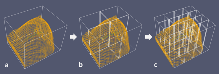
Fig. 2. Conceptual diagram of hierarchical block-wise point set matching. (a)global region, (b)2by2 blocks, (c)4by4 blocks
The result of point set matching of each block contains the information of local features, and it is used as transformation parameters for free-form deformation(FFD).
c. Transformation
The final stage is the transformation of the PET image with the optimized transformation parameters, and the fusion of the transformed PET images and the MR images. Each block has linear transformation parameters, but connected sections of blocks are to be non-linear regions. It causes being piled up or empty space. To resolve this problem, we applied B-spline based FFD.
Results
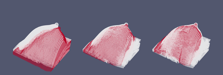
Fig. 3. The 3D rendering images of CT(red) and MR(white) surface.
Figure 3 compares the rendered images of CT and MR images. The reference MR image is displayed in gray volume while target CT image is illustrated with the red frame. Figure 3 (left) shows the image before transform. Figure 3 (middle) indicates the results of the surface matching with the conventional algorithm. Figure 3 (right) illustrates the result of our proposed algorithm, the hierarchical surface matching with modified ICP. Compared with Figure 3 (middle), Figure 3 (right) shows better matched image on the nipple. Figure 4 shows the center planes of a suspicious lesion in the fusion images after applying proposed algorithm. The MR image is displayed in gray scale, and the registered PET image is illustrated with hot color map.
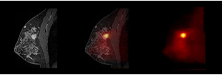
Fig. 4. Result fusion images of the proposed breast registration algorithm. The MR image is displayed in gray scale, and the registered PET image is illustrated with hot color map.
Conclusion
We have been developing free-form deformable registration algorithm for breast PET-CT and MR images based on the perturbation weighted feature information. The preliminary results show that the surface of breasts from MR and CT matched well, thereby alignment of the clinically meaningful lesions of the MR and PET slices were improved.
Participating Researchers