
ONGOING PROJECTS
- Development of Remote Radiation Detection Imaging System Mounted on a Drone
- High Resolution SPECT using Variable Pinhole Collimator
- Positioning Algorithm for CZT Virtual Frisch-grid Detector
- Multi-purpose Super-resolution Gamma Detector
- Multi-pinhole SPECT
- Low Profile Light Guide using Diffusion Film
- Advanced Reconstruction for Radiation imaging (ARRA)
COMPLETED PROJECTS
- Reconstruction of Dose Distribution in In-beam PET for Carbon Theraphy
- Image Registration for Breast Cancer Study
- High Energy Collimator Design for I-131
- Plasma-Display-Panel based X-ray Detector (PXD)
- CCD based Gamma Camera
- Abdomen Registration for PET/CT and MR images
- Performance Optimization by Patient Dose Analysis and Image Quality Assessment in CT Fluoroscopy
- Cone-beam based system matrix for a pixelated SPECT detector
- Development of Time-of-Flight Method for Improvement of Signal-to-Noise Ratio
Reconstruction of Dose Distribution in In-beam PET for Carbon Theraphy
Introduction
Positron emission tomography(PET) is has been known as an useful instrument in cancer diagnosis, localization of illness points, evaluation of treatment etc. In cancer treatment, the high energy bremsstrahlung photons in MeV scale deliver higher doses to deep-seated tumors, while reducing the doses absorbed by the surrounding healthy tissues. Beams of protons and carbon ions have a much more favorable dose-depth distribution than that of photons.
In-beam PET detects the annihilation photons which are generated during hadron therapy. The in-situ measurement allows one to acquire the maximum statistics by detecting the activity contribution by the very short half-life isotopes, and to minimize blurring effects due to patient shifts during the transport to a clinical PET system. However, the geometry of in-beam PET has one or two openings around the rings in order for the hadrons to arrive the tumor without prevention of detector blocks. The vacancy of detector modules makes truncation in projection data due to the insufficient angle coverage. Another problem is ring artifact caused by the gaps between detector modules. Gantry of PET consists of a number of detector modules, and there are gaps between each detector modules. If noise in the acquired projections is very high like in in-beam PET, these gaps may cause artifacts in reconstructed images. In this study, we aim to compensate the artifacts which come from the gaps and improve reconstruction performance using location information on the pre-determined hadron path.
Materials and Methods
A. Projection data
We acquired the list-mode data of PET whose initial ion energies are 170, 290, 350AMeV of carbon beams modeled with GATE v6.1. Each detector module consists of a 13 by 13 LYSO crystal array. The dimension of a crystal was 4by 4 by 20 mm and radius of inner circle of the gantry was 15cm. In addition the number of detector modules is 12 to 16 depending on the types of gantry: full ring, C-shaped ring, dual-head type.
B. Reconstruction of dose distribution
Low sensitivity is one of the intrinsic problems in in-beam PET. To reconstruct dose distribution from the high noise data set, we developed reconstruction method based on prior knowledge. Basic idea of the proposed method is illustrated in Fig. 1. Because most of the annihilation photons are generated along the path of carbon beam, we can approximately estimate the origin of photon pairs from the detected line of response.
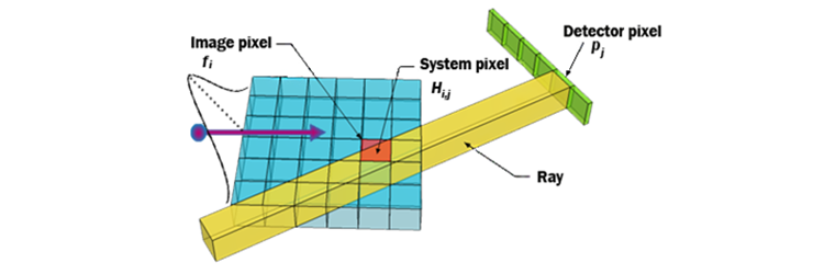
Fig. 1. Diagram of the pre-information of annihilating location.
System matrix gives the probability of detecting the photon emitted from certain point of the image at certain point of the detector.[3] Similar to time-of-flight PET reconstruction, we applied a Gaussian distribution through the width of heavy ion beams in our back-projection routine. Then expectation maximization updates were performed iteratively until it converged.
Then, the original sinogram in DCT domain was filtered by the mask in the way of element by element multiplication. After the 2-D inverse DCT transforming it, an estimate of the full sinogram data is acquired. We extract the only gap part of the full sinogram data and sum with the original sinogram. The result of this process replaced the original sinogram. This work is done iteratively.
Results and Conclusion
In Fig. 2, we compared the reconstructed dose distributions between generic OSEM and the proposed method. The Bragg peak is localized correctly while the surrounding area is represented differently.
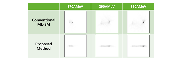
Fig. 2. The results of proposed OS-EM and General OS-EM classified into energy(C-shaped gantry type).
In addition, the results of proposed method shows that it can compensate the error caused by insufficient angle coverage. However the background value is also emphasized on path of ions. This problem is under consideration to us, and it can be suppressed before announcement. In Fig. 3, the profile shows the increased gain of data. It seems that low sensitivity of the in-beam PET can be overcome with proposed method if the background enhancement is solved.
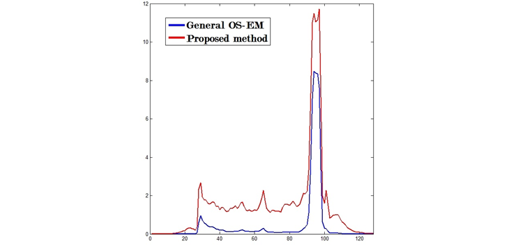
Fig. 3. Profile of the reconstruction image at midline. Initial energy of beam is 350MeV.
Participating Researchers